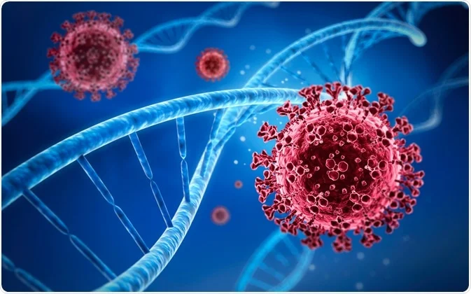
New Department of Nuclear Medicine and Molecular Imaging Technological development in molecular biology and, especially, in molecular genetics has opened up a myriad of indisputable clinical advantages in daily medical practice. The sensitivity and specificity of the techniques used in diagnosing genetic defects or etiopathogenic agents associated with morbid processes represent a valuable tool in medicine. The speed with which results are obtained must be added. For this reason, it is essential to know how DNA replicates, is transcribed and translated into proteins (central dogma of molecular biology), and the mechanisms of basal and inducible expression of genes in the genome. One of the main objectives is to know the means of regulation and gene expression in the processes of the disease to try to stop it and even prevent it,
Since the discovery that all diseases have a molecular cause, significant progress has been made in achieving effective treatment of these diseases. Any disease presents, to a greater or lesser degree, alterations in the structure, properties, metabolism or function of one or several biomolecules.
Among the causes that can induce a disease are physical agents (radiation, dust, trauma, etc.), exposure to toxic compounds (solvents, heavy metals, toxins, etc.) or biological agents (viruses, bacteria, parasites, etc.) and disorders of genetic origin whose aetiology lies in the genetic information contained in the organism.
According to the altered biomolecule, diseases can also be classified as hormonal, immunological, nutritional and metabolic, among others. Regardless of its classification, the condition is caused by cellular or tissue dysfunctions and generates temporary or permanent changes in the body as well as variations in the function of organs and systems, which are ultimately reflected in the form of illness, even those due to other causes and that is not considered to be of molecular origin.
There are different types of diagnosis of a disease according to the information analysed:
The clinical diagnosis is carried out from medical observation (exploration) with the support of laboratory data (clinical analysis, radiology and imaging, etc.). It is based on a phenotypic criterion and the manifestations under the form of the clinical syndrome.
The molecular diagnosis of a disease is based on genotypic criteria that alter the constitution of the genome. From the molecular point of view, infections can be classified into: genetic, exogenous and mixed. This classification is not widely accepted because the molecular cause of many diseases is not precisely known.
The object of study of molecular pathology is the knowledge of the disease from the point of view of its molecular alteration to contribute to its diagnosis and therapy. At the beginning of the 20th century, the expression of inborn errors of metabolism was established to describe genetic alterations present during the life of the patient, which affect specific metabolic pathways due to the absence or lack of activity of particular molecules (primarily enzymes), such as albinism, alkaptonuria, cystinuria and galactosemia.
The systematic beginning of molecular pathology took place in the 1950s, with the techniques that allow the study of human chromosomes and the knowledge of their role in sexual development and related chromosomal abnormalities.
Genomics includes the genome and the application of genetic tests to identify the alterations responsible for a disease. Similarly, transcriptomics and proteomics define the role of RNA and protein in initiating or establishing infection.
Our Department has been conceived as a comprehensive service that provides precision diagnoses within the framework of personalised attention tailored to each patient and their treating physician.
As a result of our permanent commitment to quality and medical excellence, we have inaugurated a new Department of Nuclear Medicine and Molecular Imaging SPECT / PET-CT with the most advanced technology available.
In addition, we designed an extensive and integrated area so that each patient has a comfortable and well-cared experience throughout the care process.
We are proud to have a multidisciplinary medical team led by Dr María del Carmen Alak, the leader with a recognised track record in the speciality.
Technological innovation PET-CT and SPECT (Gamma Camera)
The new PET-CT and Gamma Camera equipment, added to the scope of new radiopharmaceuticals, allow us to have a differential service regarding the diagnosis and treatment of each patient who trusts us.
- More accurate diagnoses.
- Studies in less time.
- Lower radiation rates.
- cutting edge technology
New PET-CT
We incorporated a piece of PET-CT equipment from General Electric, model DISCOVERY IQ 3.
It is a PET (Positron Emission Tomography) equipment integrated into a single Gantry with a 16-channel Multislice Computed Tomography (CT) that allows combined morphofunctional studies to be carried out.
New GE Gamma Camera
Spect double head MN 830 with Morphological Image Fusion program (CT – NMR). It responds to the “NEMA” resolution (given by the chamber of providers worldwide).
It has detectors designed to respond to the wide variety of studies carried out by Nuclear Medicine.
We care about ensuring patient safety.
Delivering low radiation rates is especially beneficial for cancer patients who often must undergo multiple studies.
All our services have radiological protection measures, guaranteeing safe studies and treatments for each patient, both adults and children.
ADVANTAGES OF THE NEW MULTIMODAL OR HYBRID PET-CT EQUIPMENT
- It allows us to see small lesions with the highest sensitivity.
- Greater comfort for the patient.
- 50% less study time.
- Quick scans of small lesions and low radiation rates.
- Morphological and metabolic detection of lesions due to its high resolution.
ADVANTAGES OF THE NEW GAMMA CAMERA
- Better image quality by reducing the radiation dose administered to the patient.
- Supports a more significant weight (up to 180 Kg. with a diameter of 60 cm).
- Reduction of study time due to the speed of exploration.
- More excellent resolution and sensitivity in the image evaluation process.
- Diagnostic and therapeutic studies.
- Cardiological studies: possibility of carrying out the protocols in one day.
- Through the administration of radiopharmaceuticals, these studies provide diagnostic information on the different metabolic processes at the cerebral, cardiological, traumatological, nephrological, pediatric and other levels.


